
MRI is the imaging modality of choice in the examination of traumatic brachial plexus injuries and thoracic outlet syndrome. MRI is useful in diagnosis and localization of these lesions. MRI is safe, non-radiative, non-invasive, having the multiplanar capability and better soft tissue characterization.
Clinical History: Right upper limb pain.
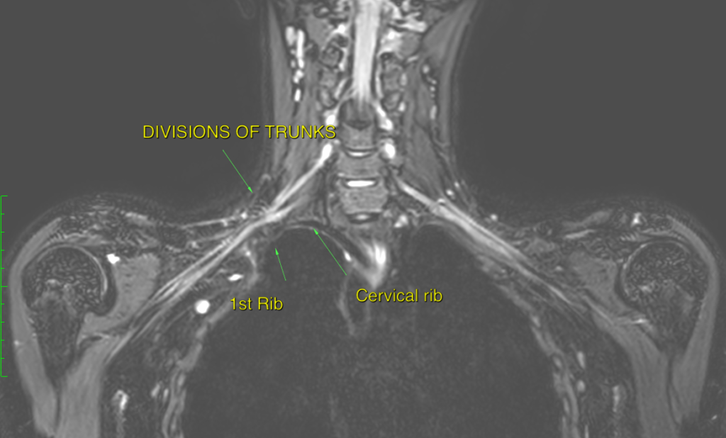
RADIOLOGICAL FINDINGS:
- Right sided complete cervical rib with pseudoarticulation with posterolateral shaft of right sided 1st rib.
- This pseudoarticulation is adjacent to divisions of right brachial plexus with indentation on divisions of inferior and middle trunks of right brachial plexus.
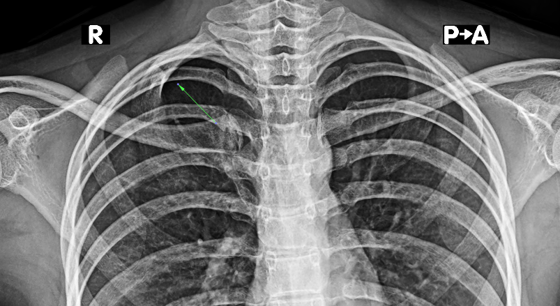
- Small incomplete cervical rib noted on left side.
Article Categories:
3.0 MRI · Case Study
Likes:
0