
WILSON’S DISEASE
3.0 T M.R.I. BRAIN
Clinical information: A 25 years old female patient having Slurred speech, both upper and lower limb Parkinson.
Findings:
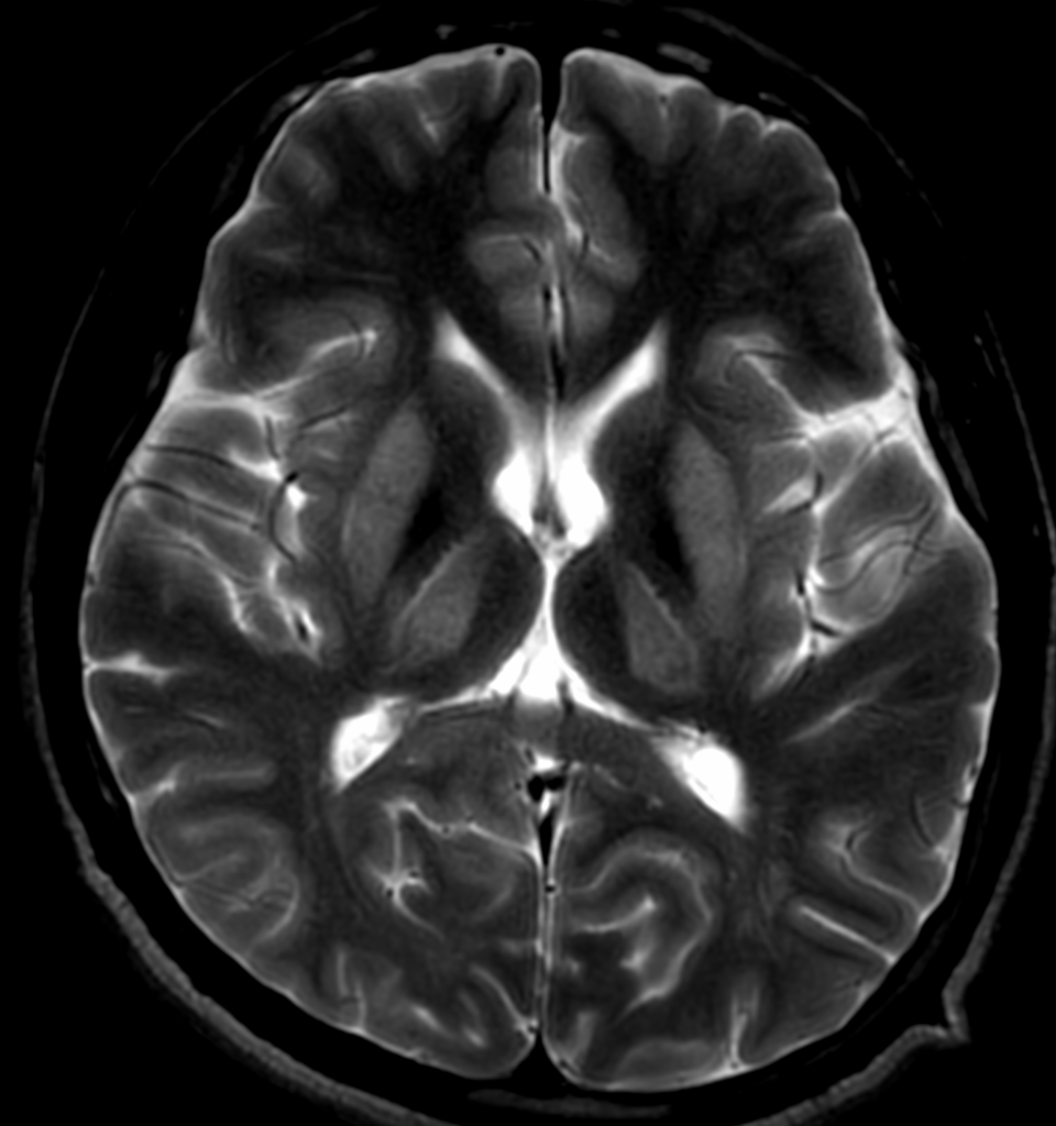
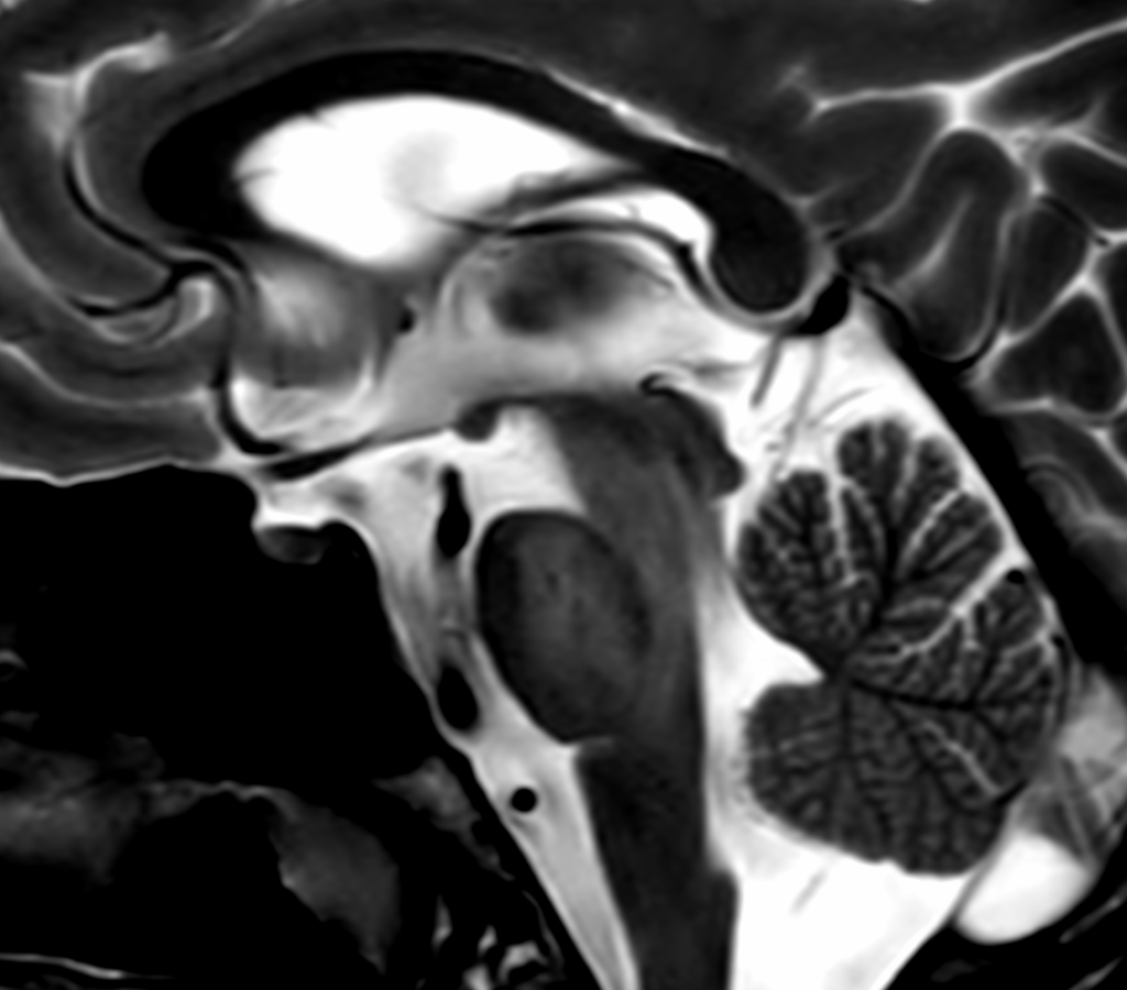
• There is symmetrical bilateral abnormal signal area in the putamen, thalami, mid brain, periaqueductal gray matter and the cerebellum (predominantly involving the dentate nuclei).
• It appears heterogeneously hyperintense on T2W and hypointense on T1W images.
• There is characteristic sparing of the red nucleus.
• Possibility of Wilson Disease.
Discussion:
• Wilson disease, also known as hepatolenticular degeneration, is a multisystem disease due to abnormal accumulation of copper.
• The deposition of copper begins immediately at birth, with symptoms usually presenting in late adolescence.
• It is characterized by early onset liver cirrhosis with CNS findings most frequently affecting the basal ganglia and midbrain.
• Ocular manifestations include Kayser-Fleischer rings in the cornea & sunflower cataracts.