
Maduramycosis (MADURA FOOT).
“DOT IN CIRCLE SIGN” ON USG & MRI.
Clinical features: Tumour like swelling (tumefaction), Discharging Sinuses, Granules.
⎯⎯⎯⎯⎯⎯⎯⎯⎯⎯⎯⎯⎯⎯⎯⎯⎯⎯⎯⎯⎯⎯⎯⎯⎯⎯⎯⎯⎯⎯⎯⎯⎯⎯⎯⎯⎯⎯⎯⎯⎯⎯⎯⎯⎯⎯⎯⎯⎯⎯⎯⎯⎯⎯⎯⎯⎯⎯⎯⎯⎯⎯⎯⎯⎯⎯⎯⎯⎯⎯⎯⎯⎯⎯⎯⎯
⎯⎯⎯⎯⎯⎯⎯⎯⎯⎯⎯⎯⎯⎯⎯⎯⎯⎯⎯⎯⎯⎯⎯⎯⎯⎯⎯⎯⎯⎯⎯⎯⎯⎯⎯⎯⎯⎯⎯⎯⎯⎯⎯⎯⎯⎯⎯⎯⎯⎯⎯⎯⎯⎯⎯⎯⎯⎯⎯⎯⎯⎯⎯⎯⎯⎯⎯⎯⎯⎯⎯⎯⎯⎯⎯⎯
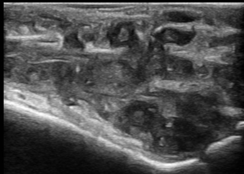
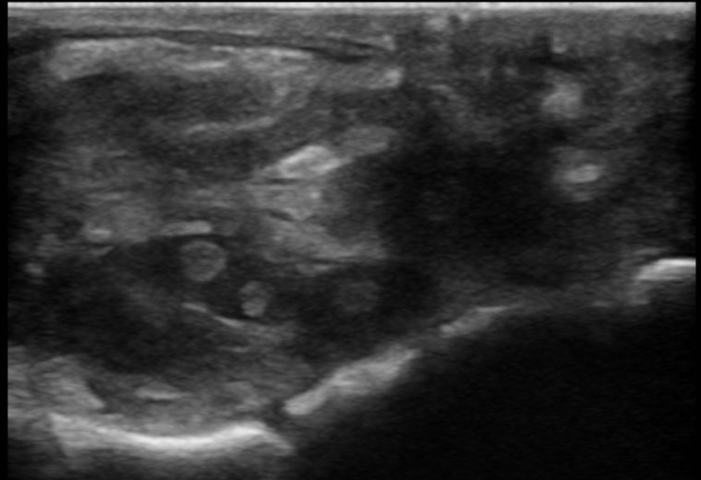
Findings (on USG):
➥ A large hypoechoic soft tissue lesion noted, It is multiloculated.
➥ It appears to be involving subcutaneous fat, vessels as well as muscle tendons.
➥ Skin sinus formation noted.
➥ “Dot in circle sign” is seen at multiple places in the lesion.
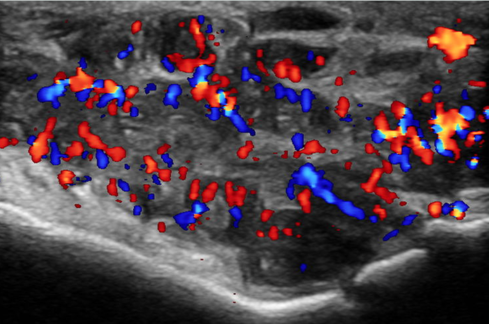
➥ Extensive vascularity is seen in the lesion on Doppler study.
➥ Few areas of liquification noted within.
Discussion:
➥ Maduromycosis, also known as maduramycosis or eumycetoma.
➥ It a chronic slowly progressive post-traumatic infection of a subcutaneous tissue usually occurring in a foot and rarely in other part of body.
➥ Caused by fungi and bacteria.
➥ A nodule, or abscess over months to years progresses to chronic infection with the formation of granulomatous nodules drained by sinuses connecting with the skin. Superimposed bacterial infection may result in larger open ulcers.
➥ MRI is modality of choice for maduramycosis and also to differentiate it from Vascular tumour being extensively vascular.